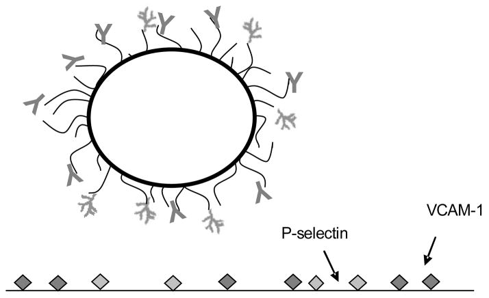
Figure 1.
Perfluorocarbon-filled microbubble targeted with MVCAM.A(429) against VCAM-1 (Y-shaped on bubble) and polymeric Slex (branch-shaped), which binds selectins. A biotin-streptavidin coupling system is used to graft biotinylated mAbs and carbohydrates on the PEG brush (black arms extending from microbubble surface) covering the phospholipid shell (thick, black circle) of ultrasound contrast agent microbubbles.

