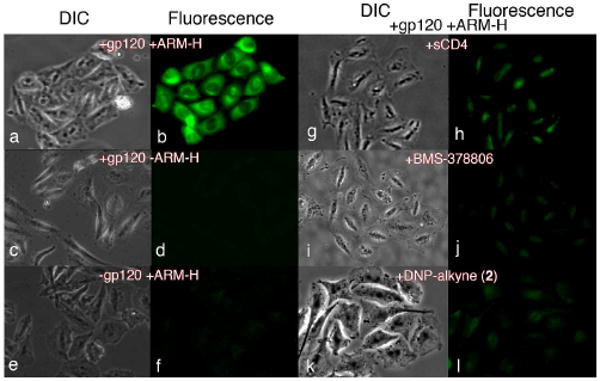
Figure 3.

Immunofluorescence Microscopy. Chinese Hamster Ovary (CHO) cells expressing HIV-1 Envelope (Env) glycoprotein (+gp120) and control cells lacking Env (-gp120) were stained with AlexaFluor488-conjugated anti-DNP antibody (15 μg/mL). Microscopy was then performed after treatment with the following reagents, as indicated: ARM-H (10 μM), sCD4 (4 μg/mL), BMS-378806 (BMS, 500 μM), or DNP-alkyne 2 (50 μM). Fluorescence signal indicates ARM-H-mediated recruitment of antibody to gp120-expressing cell.
