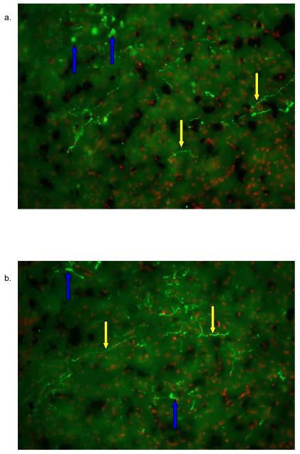
Figure 3. IF of AVP within the NAc core and shell.
AVP inmuno reactivity was detected with the NAc core (3a), and NAc shell (3b). AVP was detected with PS-41 monoclonal antibody and a goat anti mouse Cy3 (green) labeled secondary antibody. Nuclei were labeled with Hoescht dye (Red). Most of the label was found within the axons. Yellow arrows represent an axon fibers and blue arrows represent fibers that are outside the plane of focus thus showing a disperse immunofluorescence.

