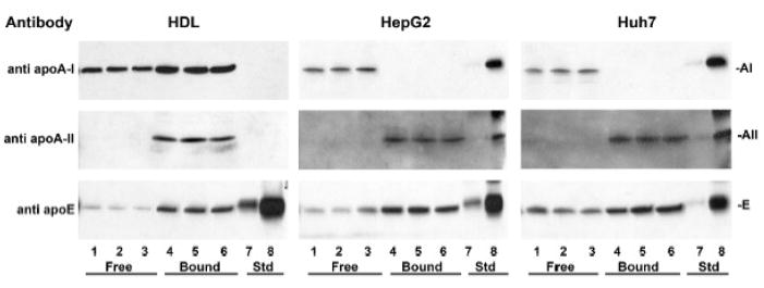
Figure 4. Intracellular apo A-I and apo A-II are on distinct particles.
Cell lysate samples from HepG2 (middle panels) and Huh7 cells (right panels) collected at 2 h were fractionated on TPS columns and probed by Western blot with antibodies to apo A-I, apo A-II and apo E. Lanes 1 – 3: Triplicate aliquots of column pass through fraction (Free). Lanes 4 – 6: Triplicate aliquots of bound fraction (Bound). Lanes 7, 8: standard apolipoproteins, 0.1 and 1 ng each, respectively. The efficiency and capacity of the TPS columns to bind reduced apo A-II and resolve LpA-I from LpA-I/A-II was demonstrated with human plasma HDL (left panels), processed in parallel with the cell lysates but loaded onto the TPS columns at more than ten times the lysate apolipoprotein concentration.

