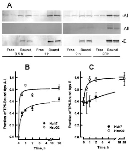
Figure 5. Time course for formation of LpAI/AII in media of hepatoma cells.
A. Western blot of TPS flow through (Free) and bound (Bound) fractions of HepG2 media collected at 0.5, 1.0, 2.0 and 20 h. Each sample was loaded in duplicate lanes. Membranes were probed with antibodies to apo A-I (top), apo A-II (middle) and apo E (bottom). Similar blots were obtained with media of Huh7 cells (data not shown). B. Rate of formation of LpA-I/A-II in HepG2 (open circles) and Huh7 (closed circles) media. C. The fraction of media apo E that binds to TPS as a function on time. Amounts of each apolipoprotein in the free and bound fractions was determined by phosphorimaging of the Western blots. The data are mean and SEM (n=4).

