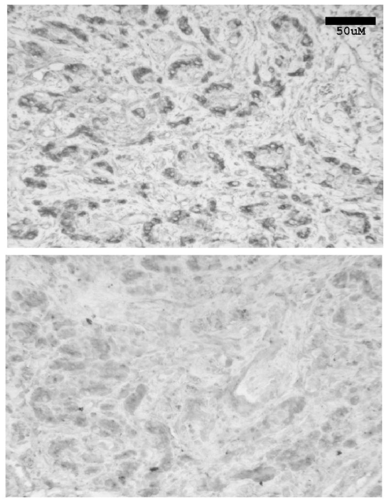
Figure 11.

Top Panel. Section of the cat CB (4-5μm thick) demonstrating immunohistochemically the presence and location of the A2A ADO receptor on the glomus cells of the cat CB. The clustering of glomus cells in glomeruli is apparent. Bottom Panel. A section 80μm removed from the positive section, the negative control showing the results of incubating a CB slice with the A2A ADO receptor antibody which had been previously incubated with the antigen. Bar: 50μm.
