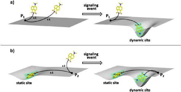
Figure 3.
(a) Attachment point P1 is located far from a hydrophobic pocket formed upon a structural change in the protein. Here, a longer linker is preferred to a shorter one. (b) Attachment point P2 is located near the hydrophobic pocket formed in response to a structural change. Such sites may favor shorter linkers as longer linkers could tend to allow the fluorophore to encounter non-specific hydrophobic patches that remain static with regard to the dynamics of protein topology. This would result in higher background fluorescence.

