
Figure 2.
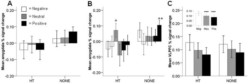
Region-of-interest % signal change. A: Amygdala % signal change did not differ between women that currently used hormone therapy (HT) and women that did not (NONE). B: Amygdala % signal change was marginally higher for early neutral scenes than for late neutral scenes in HT women (p = 0.06)*. Signal change for late positive scenes in NONE women was significantly different from zero (p = 0.02)**. First bar is early scenes; second bar is late scenes. C: Ventrolateral prefrontal cortex (VLPFC) % signal change did not differ between HT and NONE women. Inset: VLPFC signal change was higher for negative than for neutral or positive scenes (ps < 0.03)***. Neutral and positive scenes did not differ from each other.