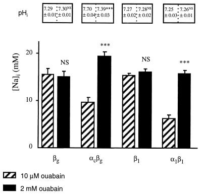
Figure 3.
Effect of 2 mM ouabain on [Na]i and pHi in X. laevis oocytes expressing various P-ATPase subunits. Oocytes were injected with various P-ATPase subunit cRNAs, as indicated below the bars. [Na]i was measured by using intracellular Na+-selective microelectrodes after a 2-day incubation of oocytes in a Ringer’s solution supplemented with 10 μM ouabain (hatched bars, from results presented in Fig. 2), or a Ringer’s solution supplemented with 2 mM ouabain (filled bars). Results are expressed as mean ± SE; n = 6–25 oocytes, from 2–6 independent experiments. Under the same experimental conditions, pHi was measured by using intracellular pH-selective microelectrodes: results are expressed as mean ± SE; n = 6–7 oocytes, from two independent experiments. Statistics were performed compared with [Na]i or pHi values measured in oocytes incubated in the Ringer’s solution supplemented with 10 μM ouabain. Statistical significance: NS, not significant; ∗∗∗, P < 0.001.

