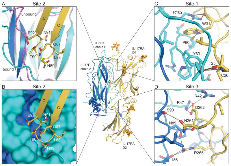
Figure 2.
IL-17F binding to IL-17RA is mediated by three distinct interfaces. (A) Site 2, the IL-17RA D1 C–C’ loop (yellow) inserts between the N-terminal coil region and strands 1 and 2 of the IL-17F chain B (cyan). The N-terminal coil undergoes a conformational change between the unbound (magenta) and bound (cyan) conformations. (B) Site 2, surface representation of the knob-in-holes IL-17F binding pocket complementarity. (C) Site 1, the IL-17RA D1 N-terminal binding site. (D) Site 3, the IL-17RA D2 binding site. Contact residues are shown as stick models. Gray dotted lines represent hydrogen bonds and pink dotted lines represent salt-bridges.

