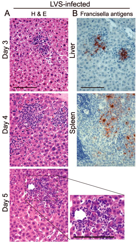
Fig. 1.

Histopathological changes in mice challenged i.n. with a minimum lethal dose of LVS (3,000 CFU) (Bokhari,et al., 2008). (A) Note that inflammatory infiltrates induced by LVS continue to grow over time with only minimum cell death and little apparent necrosis. (B) Francisella antigens are localized within inflammatory infiltrates in the liver and spleen on day 4 p.i. Bars, 100 μm.
