Fig. 2.

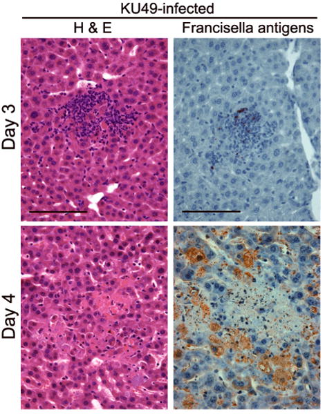
Hepatic microgranulomas form 3 days after i.n. challenge with 25 CFU of the type A F. tularensis KU49 (Wickstrum, et al., in press), and these structures are soon replaced by foci of necrosis with little inflammatory cell infiltration in the tissue parenchyma. Details of this respiratory challenge model have been published previously (Bokhari,et al., 2008). Concomitant with the death of the cells in the granulomas on day 4, bacterial antigens, detected by immunoperoxidase techniques (Bokhari,et al., 2008), become distributed throughout the liver, especially within hepatocytes, which become greatly enlarged. Bars, 100 μm.
