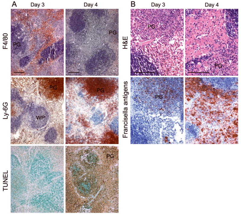
Fig. 3.

Segregated spatial distribution of cells bearing F4/80 and Ly-6G markers within the spleen of a mouse infected with the type A F. tularensis strain KU49. Infiltrating F4/80+ cells are diffusely distributed throughout the red pulp on day 3, whereas Ly-6G+ cells are predominantly localized within pyogranulomas (PG). By day 4, extensive cell death and necrosis are present throughout the red pulp, rather than the white pulp (WP). Francisella antigens are seen in both the diffuse red pulp areas and the pyogranulomas. Bars, 100 μm.
