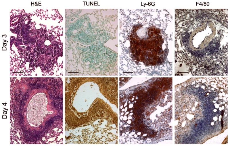
Fig. 4.

Histopathological changes in the lungs of mice infected by the i.n. route with a minimum lethal dose of type A F. tularensis KU49. Note the perivascular and peribronchial pyogranulomatous infiltrates on day 3 and day 4 and the extensive cell death (TUNEL) seen on day 4 p.i. Most of the cells in areas of cell death and necrosis bear the surface marker Ly-6G. The F4/80 cell surface marker was present on only a few infiltrating cells. Bars, 100 μm.
