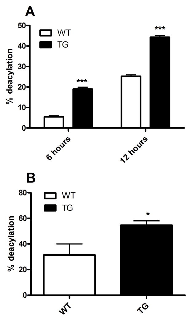
Figure 4. Rapid in vitro and in vivo LPS deacylation in transgenic mice.
(A) Thioglycollate-elicited peritoneal macrophages were incubated with radiolabeled LPS for 6 or 12 hours before deacylation was calculated as described in Methods. Percent deacylation = percent loss of secondary acyl chains. Equivalent numbers of wildtype and transgenic mouse macrophages were used (~0.15 mg protein/well). (B) 5μg radiolabeled LPS were injected intravenously into mice, livers were harvested 4 hours later, and LPS deacylation was determined as described in Methods. n = 4 mice/group. Data are representative of 2 independent experiments. For comparisons between wildtype and transgenic macrophages at each time point: * p<0.05, *** p < 0.001 (Student’s t-test). WT = wild-type; TG = AOAH transgenic.

