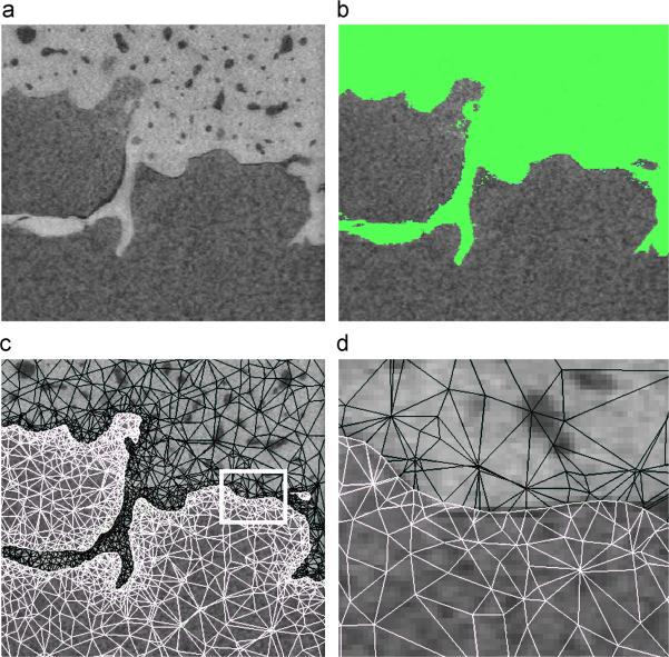
Figure 2.
a) μCT-data of the cement-bone interface.
b) Segmentation of the μCT-data into bone (1,000 to 3,071) and cavities (−1,024 to 100). Remaining gaps in the bone were filled manually.
c) Solid mesh of the bone and cement plotted on top of the μCT-data.
d) Zoomed node-to-node interface from figure c.

