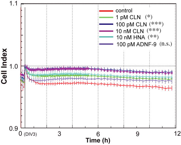
Figure 2.
Effect of colivelin (CLN), C8A-humanin, and ADNF-9 against alcohol exposure in primary cortical neurons (PCNs). PCNs were treated with or without the indicated concentrations of peptides along with ALC. Cell index, which reflects cell viability, measured by xCELLigence system is shown as mean ± SD (n=6). Cell index of PCNs without alcohol exposure is equal to 1.0 [the baseline]. Control (water), red; 1 pM CLN, light green; 100 pM CLN, navy; 10 nM CLN, purple; 10 nM C8A-humanin (HNA), light blue; and 100 pM ADNF-9, thin purple. The one-way ANOVA at 8 hours revealed a significant difference between groups (F(5,35)=7.940 [p<0.0001]). * p<0.05, ** p<0.01, *** p<0.001, n.s. means no significant difference (Dunnett’s post hoc test).

