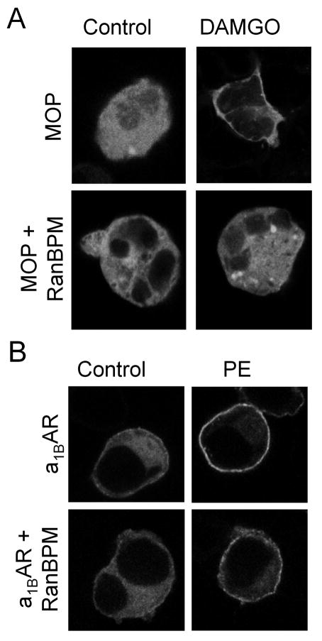
Figure 4.
MOP-stimulated translocation of βarr2-GFP is selectively inhibited by RanBPM. (A) Agonist-mediated recruitment of βarr2-GFP was visualized by confocal microscopy of HEK293 cells transiently transfected with βarr2-GFP and FLAG-MOP without (MOP) and with RanBPM (MOP/RanBPM). Cells were treated with either 10 μM DAMGO or vehicle for 10 min. (B) Agonist-mediated recruitment of βarr2-GFP was visualized by confocal microscopy of HEK293 cells transiently transfected with βarr2-GFP and FLAG-α1BAR without (α1BAR) and with RanBPM (α1BAR/RanBPM). Cells were treated with either 1 μM phenylephrine (PE) or vehicle for 10 min. Agonist stimulation resulted in the redistribution of βarr2-GFP from the cytosol to the plasma membrane in both MOP and α1BAR cells (panels A & B). No translocation of βarr2-GFP was observed following MOP-stimulation in cells over-expressing RanBPM (panel A, DAMGO), whereas α1BAR-mediated βarr2-GFP translocation was not altered by RanBPM over-expression (panel B, PE).

