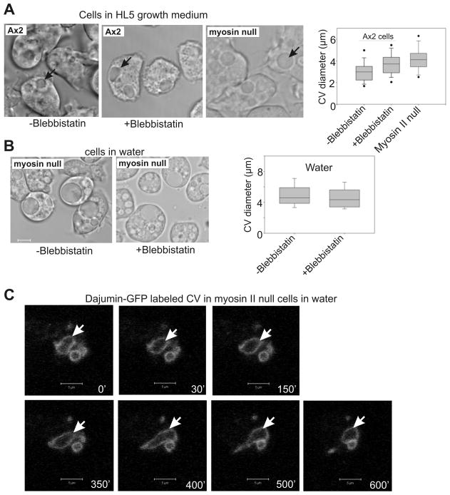
Figure 7. Myosin II is necessary for normal contractile vacuole function.
(A) Parental Ax2 cells display enlarged CVs in the presence of blebbistatin. Myosin II null cells also display enlarged CV diameters in HL5 medium. Box and whisker graph quantifies average maximal CV diameters, determined from video analysis. Average maximum CV size for Ax2 cells in blebbistatin, and myosin II null cells, both differ from the untreated Ax2, P=<0.02, (n=53–54). (B) Myosin II null cells display larger maximum CVs in water than do parental Ax2 cells, and size is not affected by the myosin II inhibitor blebbistatin. (C) Myosin II null cells expressing CV marker protein dajumin-GFP display impaired CV dynamics. Cells were imaged after transferring cells from HL5 medium to water. Arrow denotes a persistent contractile vacuole. Scale Bars, 5 um. See movie 4, CV dynamics of parental Ax2 cells in water, no blebbistatin; movie 5, CV dynamics of Ax2 cells in water, with blebbistatin; movie 6, CV dynamics of myosin II null cells in water, no blebbistatin; movie 7, CV dynamics of myosin null cells in water, with blebbistatin.

