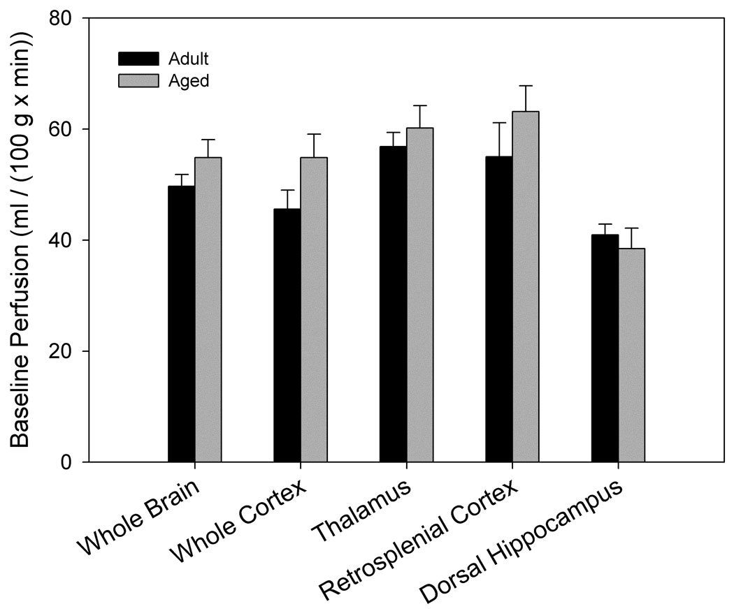
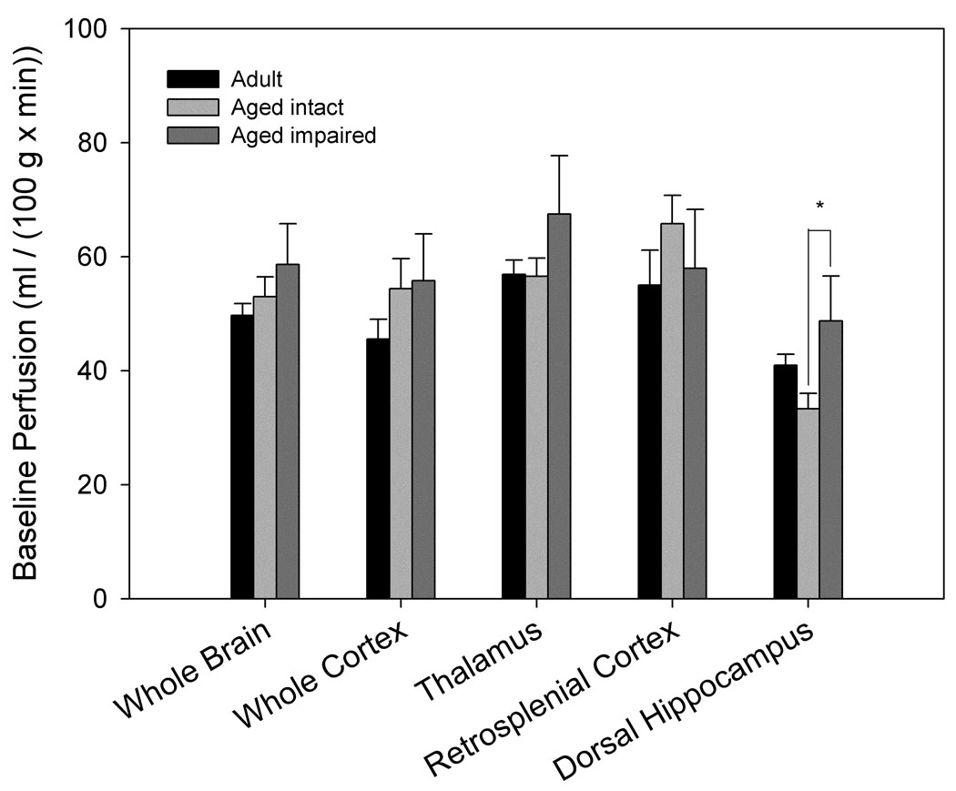
Figure 2. Basal cerebrovascular perfusion differs by spatial memory performance.
High resolution images using a rapid acquisition, relaxation-enhanced (RARE) pulse sequence were acquired for morphologic reference purposes. Regions of interest were manually outlined and included the entire cross-section of the brain, cortex, dorsal hippocampus, retrosplenial cortex and the thalamus. For FAIR analysis, the recovery curves obtained from each pixel of the global or selective inversion images were numerically fitted to an exponential model and longitudinal recovery rates were then used to calculate the perfusion rate, f (ml/(100g × min)) on a pixelwise basis using the relationship f = λ · (1/τNS−1/τSS). Areas overlapping large blood vessels or ventricles were removed. A. No significant differences in basal perfusion were found between age-groups (nadult= 6, naged= 12). B. Separation of aged animals by maze performance revealed significantly higher perfusion of dorsal hippocampus in aged impaired (n= 4) when compared to aged intact (n= 8) animals (p= 0.045).


