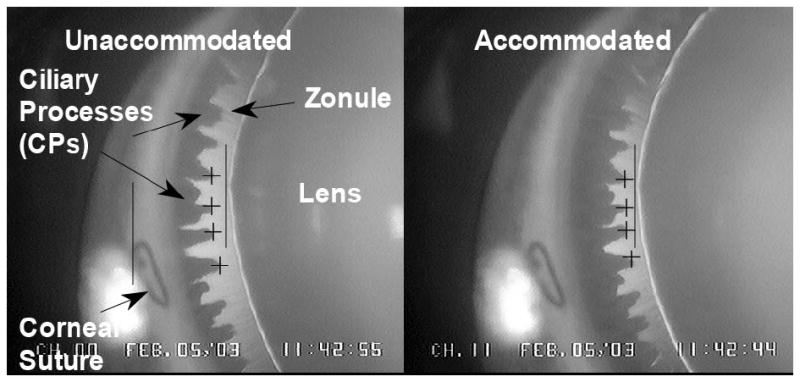
Figure 1.

Goniovideography images of normal lens and ciliary process (CP) configuration in the accommodated and unaccommodated states. To obtain quantitative measurements, a 9-0 nylon suture placed at the corneoscleral limbus served as a reference point (left solid vertical line) from which to measure distances to the lens equator (right solid vertical line) and the CPs (cross-hairs) for each image during a 2.2-sec stimulus period. Reprinted with permission from: Croft et al. Accommodative Ciliary Body and Lens Function in Rhesus Monkeys I. Normal lens, zonule and ciliary process configuration in the iridectomized eye. Invest Ophthalmol Vis Sci 2006;47:1076-1086.
