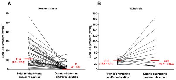
Figure 3.
Nadir LES pressure during periods of distal esophageal shortening and/or prolonged LES relaxation in non-achalasic patients (Panel A) and achalasia patients (Panel B). Median (5th - 95th) percentiles are indicated in red. In each case, nadir LES pressure during the event is compared to the lowest pressure during a comparable time period prior. Most (57/64) non-achalasia patients had LES pressure nadirs to ≤4 mmHg qualifying the event as a complete tLESR. On the other hand none of the fifteen achalasia patients had an LES pressure nadir ≤ 4 mmHg and half of them actually had significantly increased LES pressure during the shortening event. Note that the pressure scales of the two plots are different.

