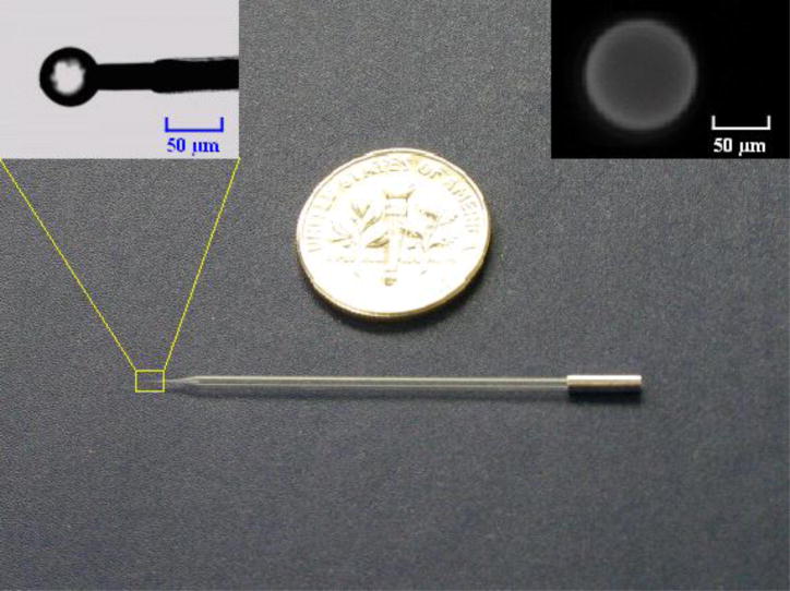
Figure 1.
A photograph of the agarose bead-based micropipette biosensor shown next to a US quarter coin. The left inset is an enlargement of the probe’s tip, showing the dry, functionalized agarose bead mounted on the platinum electrode. The right inset shows a fluorescent image of the hydrated biotin agarose bead labeled with streptavidin-Alexa Fluor 488 and suspended in PBS solution.

