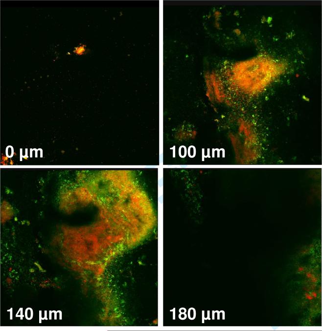
Figure 1.
Confocal images (horizontal X-Y sections) obtained by dental-plaque derived biofilms grown on agar in 24-well plates. Live bacteria with intact membranes were stained fluorescent green by SYTO 9 stain, while dead bacteria with damaged membranes were stained fluorescent orange by propidium iodide. The fluorescent signals were obtained to a depth of 180 μm.

