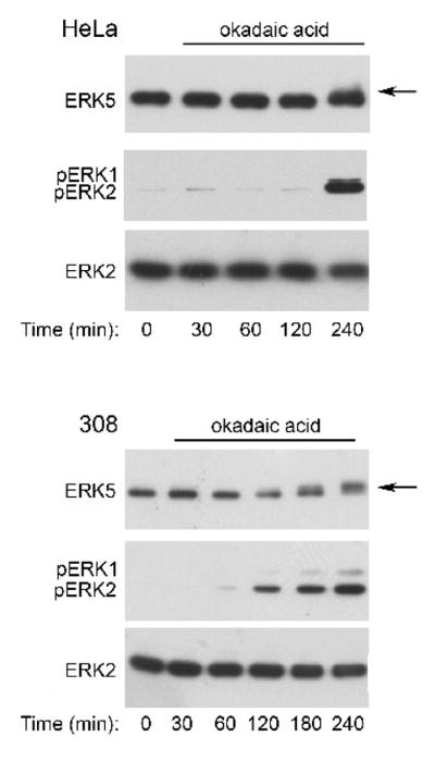
Figure 9. Okadaic acid stimulates ERK5 phosphorylation with different kinetics than palytoxin in 308 cells.

HeLa and 308 cells were incubated for the indicated times with 120 nM okadaic acid. Protein (20 μg) from whole cell lysates was analyzed by immunoblot for the following: ERK5; phosphorylated, active ERK1/2 (pERK1 and pERK2); and total ERK2. The arrow marks the slower migrating form of ERK5. The data shown are representative of at least two independent experiments.
