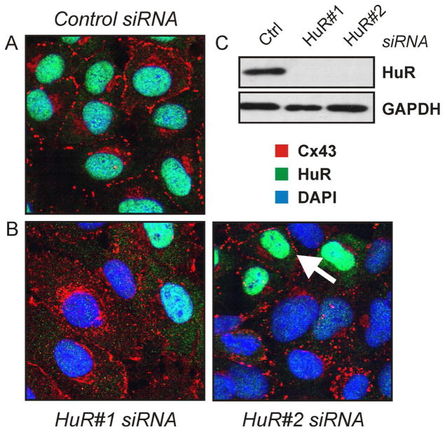
Figure 3.
Subcellular localization of connexin-43 in cells depleted of HuR. (A, B) WB-F344 cells were treated with control or one of two different HuR-specific siRNAs for 48 h, followed by immunocytochemical analysis by confocal microscopy of Cx43 (red) and HuR (green) localization. Nuclei were stained with DAPI (blue). The arrow indicates a cell with insufficiently depleted HuR displaying Cx43 localization similar to control cell conditions. (C) Western analysis of HuR levels in cells treated with control or HuR-specific siRNAs. GAPDH levels were analysed after treatment with control and HuR siRNA. Images are representative of at least three independent experiments

