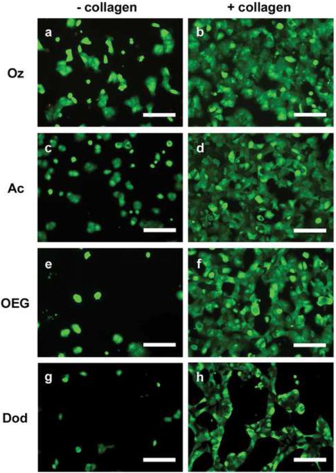
Figure 4.
Representative optical micrographs of primary rat hepatocytes seeded on 2 types of chemically modified porous Si samples with (+ collagen) and without (− collagen) adsorbed collagen type I. Abbreviations for the samples are indicated on the left: tissue culture polystyrene (PS), ozone-oxidized (Oz), undecanoic acid (Ac), oligo(ethylene) glycol (OEG), and dodecyl (Dod). Cells are stained with the vital dyes calcein acetoxymethyl and ethidium homodimer I. (a) Hepatocytes on ozone-oxidized porous Si. (b) Hepatocytes on ozone-oxidized porous Si pretreated with collagen. (c) Hepatocytes on undecanoic acid-terminated porous Si. (d) Hepatocytes on undecanoic acid-terminated porous Si pretreated with collagen. (e) Hepatocytes on OEG-modified porous Si. (f) Hepatocytes on OEG-modified porous Si pretreated with collagen. (g) Hepatocytes on dodecyl-terminated porous Si. (h) Hepatocytes on dodecyl-terminated porous Si pretreated with collagen. Scale bar is 100 μm.

