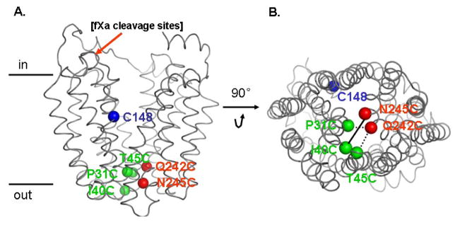
Fig. 1.
Position of Cys148 and paired-Cys replacements on the backbone of the X-ray crystal structure of LacY (PDB ID 2V8N). Left: side view; Cys148 (blue sphere; helix V); pairs are Ile40→Cys (green sphere; loop I/II) with Asn245→Cys (red sphere; helix VII); Pro31→Cys (green sphere; helix I) with Gln242→Cys (red sphere; helix VII) and Thr45→Cys (green sphere; helix II) with Gln242→Cys (red sphere; helix VII). Tandem fXa protease sites inserted between residue 136 and 137 are also shown in black. Right: viewed from the cytoplasm.

