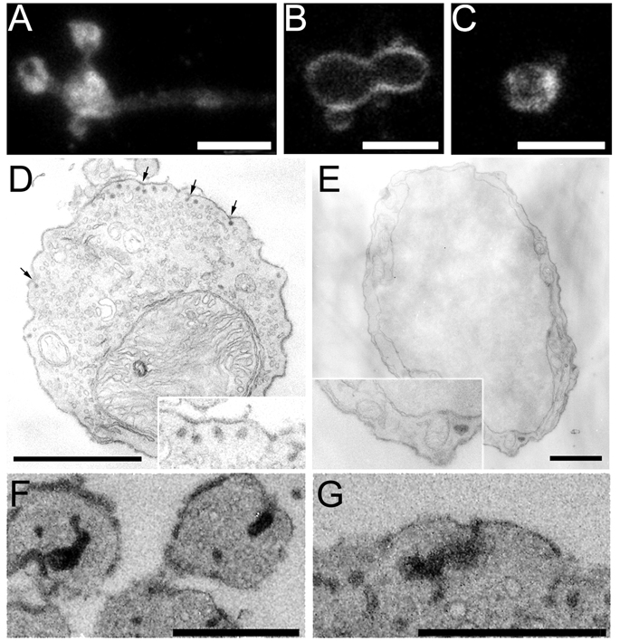
Figure 6.
Membrane retrieval is modified when dynamin GTPase is inhibited by dynasore. A, Confocal image of an isolated mouse bipolar cell terminal immunolabeled with an antibody against dynamin-1. B, C, Confocal images of terminals briefly stimulated by high K+ in the presence of FM4-64. Control cells were bathed in buffer containing 0.16% DMSO (C) and experimental cells were bathed in buffer containing 80 µM dynasore dissolved in 0.16% DMSO (B). Images were taken ~5.5 min after stimulation. Dye appears to be trapped at the membrane in cells exposed to dynasore, while control cells internalized the dye. Scale bars in A–C represent 5.0 µm. D, EM image of a cell exposed to 80 µM dynasore and stimulated with a 3-s puff of high K+ in the presence of AM1-43. Cell was fixed ~ 3.0 min after stimulation and subsequently photoconverted. Small vesicles containing photoconverted AM1-43 appear “stuck” on the membrane (arrows). Scale bar represents 1 µm. Inset shows a higher-magnification view of some of the labeled structures. E, The cell body of the same bipolar neurons shown in A lacks structures containing photoconverted AM1-43. Scale bar = 1 µm. F, G, Large structures containing photoconverted AM1-43 appear to be attached to the plasma membrane. Scale bars represent 0.5 µm.

