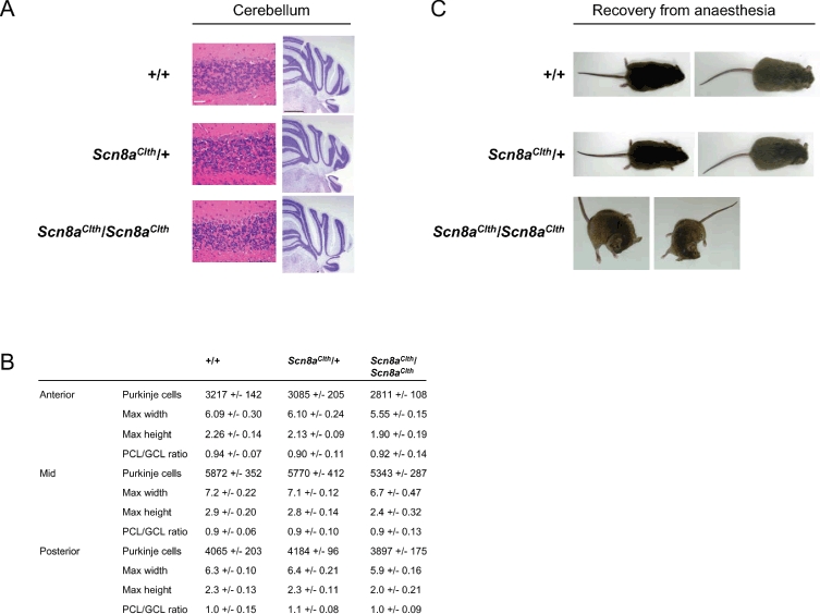
Figure 4:

(a) H&E-stained coronal sections of cerebellum from 8-month-old mice show no Purkinje cell loss in any region (scale bar, 50 μm). Histopathology of the cerebellum of 8.5-month-old mice using cresyl violet on coronal sections shows no neuronal mislocalization (scale bar, 1 mm). (b) Area and layer measurements and Purkinje cell counts of Scn8aClth mice. Although Scn8aClth/Scn8aClth cerebellums are slightly smaller and show slightly reduced Purkinje cell counts compared with +/+ and Scn8aClth/+, the Purkinje cell layer (PCL) to granule cell layer (GCL) ratio is normal, indicating normal overall cerebellar structure. Area measurements are in millimetres. (c) Abnormal dystonic postures displayed by Scn8aClth/Scn8aClth mice during recovery from anaesthesia were not seen in +/+ or Scn8aClth/+ mice. Postures were maintained for up to 1 min.
