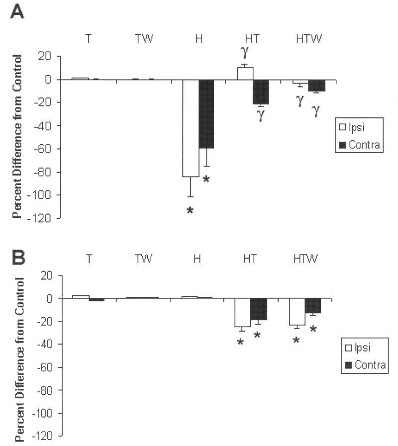
Figure 6.
A. In noninjured animals administered theophylline with or without interruption (T,TW) adenosine A1 receptor immunoreactivity was unaltered compared with naïve noninjured animals. However, C2HS rats (H) demonstrated a significant (* P<0.05) decrease in protein expression (number of labeled cells) compared with noninjured animals (the decrease was apparent ipsilateral as well as contralateral to injury). Although the ipsilateral decrease appeared more pronounced than contralateral, this was not significant. After a 3-day administration of theophylline (HT) the decrease in protein levels was significantly attenuated (γ P< 0.05 on the ipsilateral and contralateral sides. When theophylline was withdrawn for 12 days prior to assessment (HTW), the number of A1 immuno-reactive cells was still significantly lower compared with H (injury alone). (B). In marked contrast to the decrease in A1 protein levels after C2 hemisection, A2A protein levels were not altered compared with controls (T, TW) and with naïve, noninjured animals. However, after theophylline administration, protein levels significantly decreased (P<0.05) ipsilateral as well as contralateral to injury. Although the decrease appeared more pronounced ipsilaterally, (a trend similar to that observed for A1 as shown above) it was not significant. Withdrawal of theophylline for 12 days prior to assessment did not alter the decrease in protein levels, i.e., theophylline-induced decrease in A2A protein levels persisted. *=significant from control, γ= significant from hemisection.

