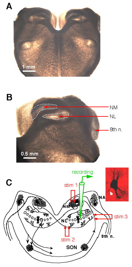
Figure 1.

Experimental preparation. A and B: Photographs (bright field illumination) of a chicken brainstem slice (300 μm in thickness). No staining was performed. The stump of the 8th nerve, the nucleus magnocellularis (NM) and nucleus laminaris (NL) can be easily identified. C: A schematic diagram showing the time-coding circuits in the avian auditory brainstem. The auditory nerve innervates the cochlear nucleus angularis (NA) and the NM. Neurons in the NM send bilaterally segregated excitatory projections to the NL. Stimulation electrode 1 (stim 1) was placed in the ipsilateral NM to activate the dorsal excitatory input to the NL, and stim 2 on the fibers ventral and medial to the NL to activate the excitatory input from the contralateral NM. Neurons in the NL receive GABAergic inputs primarily from the ipsilateral superior olivary nucleus (SON). Stim 3 was placed on the SON fibers to activate the GABAergic pathway. The inset shows the morphology of one NL neuron revealed by biocytin staining (axon indicated by the white arrow).
