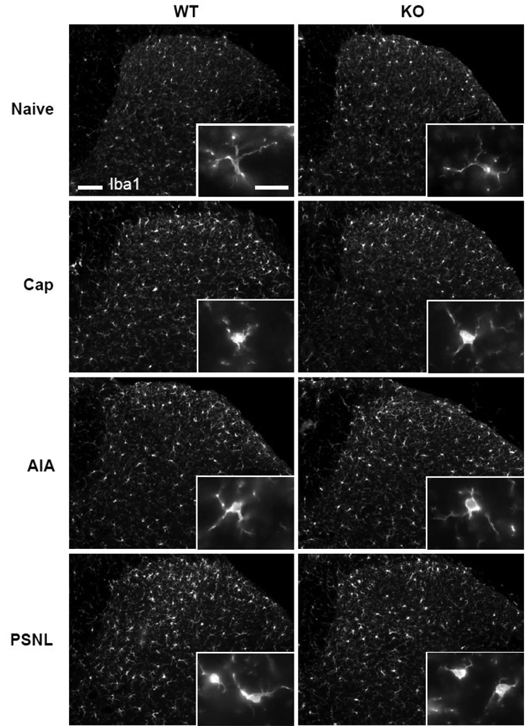
Fig. 2.
Immunostaining for Iba1 in naïve wild type (WT) and TRPV1-KO (KO) mice and in pain models (the sections are from the dorsal horn of spinal segment L4 ipsilateral to the treatment: Cap, 3 days after intraplantar capsaicin; AIA, 14 days after intraarticular CFA; PSNL, 14 days after partial sciatic nerve ligature). Enlargements in the insets demonstrate the changes in cell shape of microglia between naïve mice and various treatments. Scale bars, 100 µm and 20 µm in the inset.

