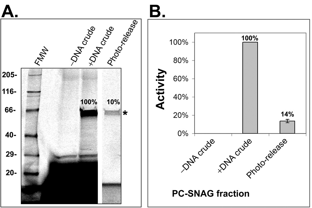
Figure 2. Analysis of PC-SNAG based on SDS-PAGE and a luciferase functional activity assay.
Luciferase was cell-free expressed and TRAMPE labeled using PC-biotin and BODIPY-FL misaminoacylated tRNAs together. PC-SNAG was achieved based on the directly incorporated PC-biotins. (A.) Fractions were separated by SDS-PAGE and luciferase (asterisk) imaged via the TRAMPE incorporated BODIPY-FL labels. (B.) Functional activity of luciferase was measured using a chemiluminescent substrate based assay. FMW = fluorescent molecular weight standards; –DNA Crude = Crude unprocessed cell-free expression reaction performed without the expressible luciferase DNA; +DNA Crude = Crude unprocessed cell-free expression reaction performed with the luciferase DNA (.i.e. actual total starting luciferase); Photo-Release = The photo-released luciferase fraction obtained by PC-SNAG. The photo-released luciferase is expressed as a percent of the “+DNA Crude” luciferase.

