
Figure 5.
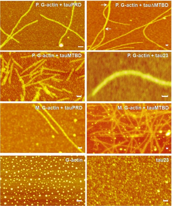
Interactions of tauPRD with G-actin observed by atomic force microscopy. Skeletal muscle (M) and platelet (P) G-actin were incubated with tauPRD in binding buffer at 37°C for 30 min and then aliquots were taken for observation by atomic force microscopy as indicated. Bars in panels are 50 nm.