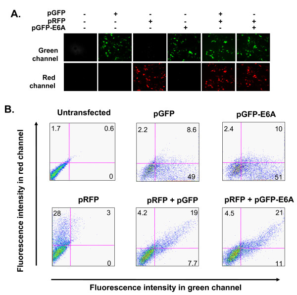
Figure 2.
Verification of splicing pattern ofrom transfected pGFP-E6A reporter minigene. A. HEK293 cells were transfected with combinations of pGFP, pRFP and pGFP-E6A as shown above each pair of photographs. After 48 hr, the fluorescence was assessed in the green and red channels. All photographs taken with the same exposure to compare intensities. B. Flow cytometry of HEK293 cells 48 hr post transfection assessing fluorescent intensity in red and green channels. The percentages of fluorescent cells are shown in each quadrant.

