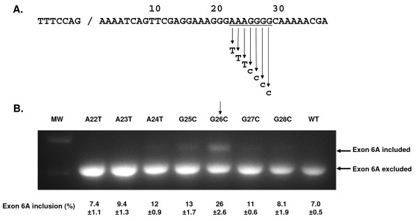
Figure 5.
Effect of point mutations within the VEGF exon 6A putative exonic splicing silencer. A. Partial sequence of VEGF exon 6A with splice donor site and putative ESS sequence (underlined). The seven mutant versions with purine to pyrimidine transversions are shown. B. RT-PCR analysis of splicing pattern 48 hr following transfection of pGFP-E6A single nucleotide substitution plasmids into HEK293 cell. The anticipated positions of the products for the splicing events with inclusion or exclusion of exon 6A are indicated by arrows. MW = lane with molecular weight markers. The exon inclusion percentage was calculated following densitometry of the gel as [E6A included band/(E6A included band + E6A excluded band)] × 100. Experiment was repeated 3 times and the average % inclusion from 3 replicate transfections and the standard error was shown.

