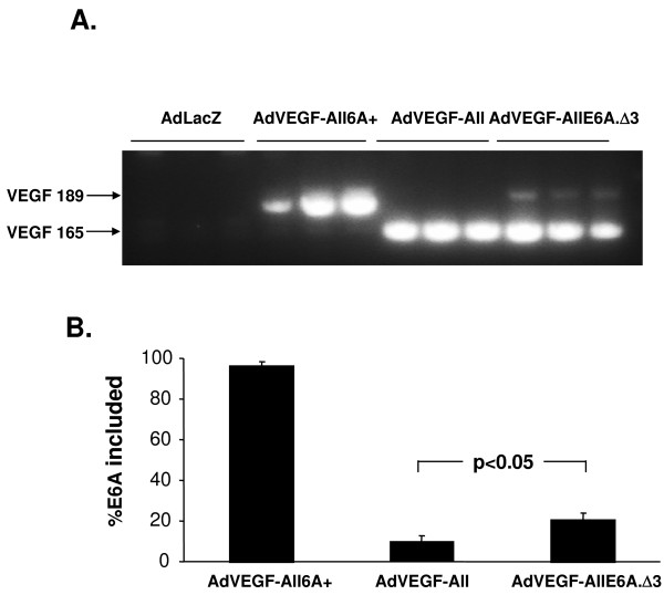
Figure 7.
In vivo effects of deletion of exon 6A nucleotides 22-30 on splicing in genomic context. Mice (n = 3/group) were administered 1010 particle units of AdlacZ as negative control, AdVEGF-All, AdVEGF-All6A+ or AdVEGF-AllE6A.Δ3 by injection into the tail vein. After 2 days the mice were sacrificed and the livers were harvested. Total RNA of each liver sample was treated with DNase I. A. One-step RT-PCR analysis of splicing pattern. Each lane represents RT-PCR product from a different mouse. The anticipated positions of the products for the splicing events with inclusion or exclusion of exon 6A are indicated by arrows. MW = lane with molecular weight markers. B. The Exon6A inclusion percentage was calculated from densitometric scan of gels as described for Figure 4, and the mean ± standard deviation is plotted for each group.

