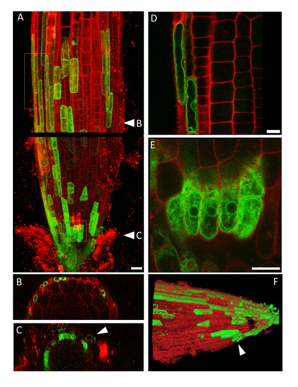
Figure 3.
YFP-TIP1;2 is expressed in the root cap. 8-day old roots from the indicated transgenic lines were excised, stained with propidium iodide for 2 min and visualised by CLSM. Stacks of 80 optical z sections (1 μm step-size) were collected from root tips. The images show a representative result for this construct. The signals from YFP fluorescence (green) and propidium iodide fluorescence (red) are merged. A: maximal 3D projection of the root tip at the base of the elongation zone. The image shows two adjacent z-stacks of the same root, separated by a black line. B and C: xz projections of the image stack in panel a, revealing two cross-sections of the root axis, taken in the regions of the root indicated by the arrowheads in A. D and E: the regions indicated by dotted boxes in A were observed at high magnification. Single optical sections are shown. Note YFP-TIP1;2 in the ER of young root cap cells and in the tonoplast of root cap cells closer to the elongation zone. F: The fluorescent traces from YFP (green) and propidium iodide (red) from the image stack in panels A were reconstructed, segmented and rendered in 3D with Mimics 12.1. Scale bars: (a), 20 μm; (d) and (e), 10 μm.

