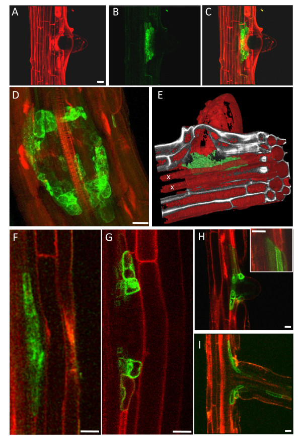
Figure 4.
TIP 2;1-YFP expression in lateral root primordia. A-E: 8-day old roots from TIP2;1-YFP transgenic seedlings were excised, stained with propidium iodide and visualised by CLSM. Stacks of 80 optical z sections (1 μm step-size) were collected from mature root axes. The images show representative results for this construct. Maximal projections of the z-stacks are shown, with the individual signals for YFP (A), propidium iodide (B) or the merged signals (C and D). E: The fluorescent traces from YFP (green) and propidium iodide (red) from the image stack in panels (A-C) was reconstructed, segmented and rendered in 3D with Mimics 12.1. Note that the TIP2;1-YFP-expressing cells are in close proximity to the xylem (labelled with x). F-I: Roots from 8-day old transgenic seedlings expressing TIP2;1-YFP (green) and TIP2;3-RFP (red) were imaged. Sequential stages of lateral root development are shown. Inset in H: note the boundary between pericycle cells expressing TIP2;3-RFP (top) and TIP2;1-YFP (bottom). Scale bars: 20 μm.

