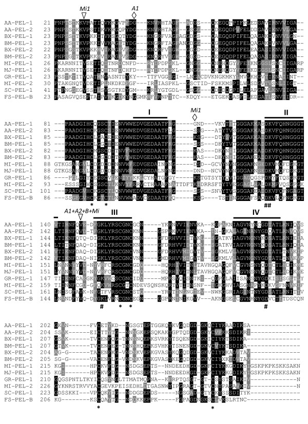
Figure 7.
Multiple sequence alignment of A. avenae pectate lyase protein sequences (AA-PEL-1 and AA-PEL-2) with the sequences of pectate lyases from plant parasitic nematodes, bacterium, and a fungus. BX-PEL-1 [BAE48369], BX-PEL-2 [BAE48370] from Bursaphelenchus xylophilus; BM-PEL-1 [BAE48373], and BM-PEL-2 [BAE48375] from Bursaphelenchus mucronatus; MI-PEL-1 [AAQ09004] and MI-PEL-2 [AAQ97032] from Meloidogyne incognita; MJ-PEL-1 [AAL66022] from Meloidogyne javanica; GR-PEL-1 [AAF80747] from Globodera rostochiensis; SC-PEL-1 [NP625403] from Streptomyces coelicolor and FS-PEL-B [AAA8738] from Fusarium solani. Identical residues are highlighted in black and functionally conserved are in gray. Black bars (I to IV) indicate the conserved regions characteristic of PL3 pectate lyases. Very highly conserved charged residues are indicated beneath the alignment by a number symbol (#), while an asterisk (*) indicates conserved cysteine residues. The positions of the intron in AA-PEL-1 and AA-PEL-2 are indicated by A1 and A2 respectively, that in BX-PEL-1/2 and BM-PEL-1/2 by B, and those in MI-ENG-1 by Mi1. Triangles and diamonds represent phase 0 and 1 introns, respectively.

