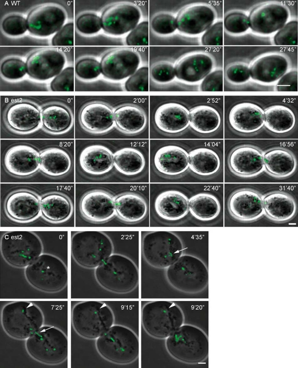
Figure 3.

Time-lapse imaging of the movement of five lacO-tagged telomeres during mitosis. One optical section is shown per time point. (A) Telomerase-positive yKS58 yeast cell in mitosis. The telomeres move one by one from the mother cell into the daughter cell. (B) Senescence/crisis in telomerase-negative yeast cells derived from yKS58. During senescence/crisis the telomeres move between both cells without separation. Mother and daughter cell are equal in size and larger than WT cells. (C) Telomerase-negative yeast cell during senescence/crisis showing that most telomere foci move between cells but at least one telomere (arrowhead) and possibly a second telomere (star) stayed positioned away from the bud neck. Most foci move through the bud neck as a dot but in one case the signal is elongated (arrow). Bar = 2 µm.
