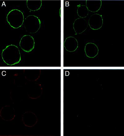
Figure 3.
Localization of the γ subunit in infected Sf-9 cells. Confocal microscopy was used to determine the location of proteins in cells infected with γ or α1β1γ baculoviruses. (A) Cells infected with γ baculovirus and probed with G17 polyclonal. (B–D) Cells coinfected with α1β1γ baculoviruses and probed with both the G17 polyclonal and 5α monoclonal to detect the γ and α subunits. (B) Staining for the γ subunit. (C) Staining for the α1 subunit. (D) an overlay of B and C, showing colocalization of γ and α1 in α1β1γ-infected cells.

