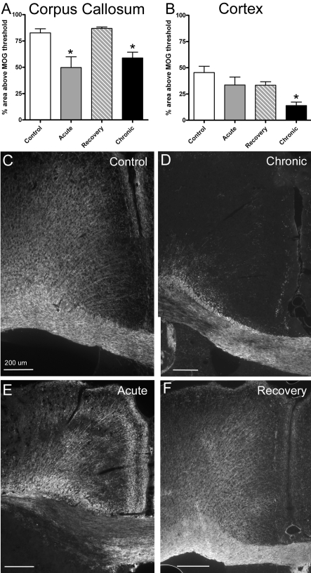
Figure 3. Quantitative analysis of myelination following behavioural assessments.
CC (A) and cerebral cortical (B) myelination was estimated in coronal brain sections based on the percentage area immunolabelled for MOG. The region analysed for the CC extended from the midline to under the apex of the cingulum. The region analysed in the adjacent cerebral cortex was outlined from the midline along the pial surface with the lateral border parallel to the midline down to the apex of the cingulum and continuing with the inferior boundary as the border with the cingulum and CC. (A) Compared with non-treated control mice, during cuprizone treatment myelination of the CC was significantly reduced at acute (6 weeks; P = 0.0027) and chronic (12 weeks; P = 0.0027) time points, whereas the percentage area myelinated returned to control levels in the mice allowed a recovery period (6 weeks of cuprizone feeding followed by 6 weeks on normal chow). (B) Compared with non-treated mice, only the chronic (12 week) cuprizone condition showed a significant reduction in the percentage area myelinated in the adjacent cortical region (P = 0.0207). (C and D) Representative images of immunofluorescence for MOG to illustrate myelination patterns in the CC and adjacent cortex in coronal sections. Comparison with a non-treated mouse (C, Control) shows demyelinated areas in both the CC and adjacent cortex after 12 weeks of cuprizone treatment (D, Chronic). After cuprizone treatment for 6 weeks (E, Acute) demyelination is much more apparent in the CC than in the adjacent cortex and the CC is no longer markedly demyelinated after a subsequent 6 week period on normal chow (F, Recovery). Scale bar = 200 μm.

