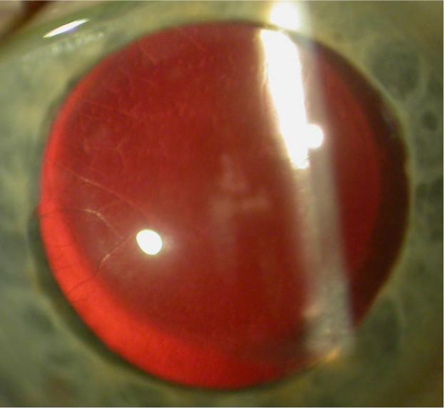
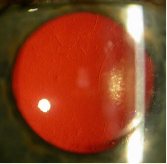
Figure.


Slit lamp photomicrographs of a 65-year-old man with thin, branching opacities seen in retroillumination against the red reflex in the right (right image) and left (left image) corneas. Focal, non-linear, opacities that also appear translucent on retroillumination are present in the central and mid-peripheral regions of each cornea.
