
Figure 5. Macrophage-like cells inhibit class switching by trafficking membrane and vesicular Nef via Vav and small GTPases.
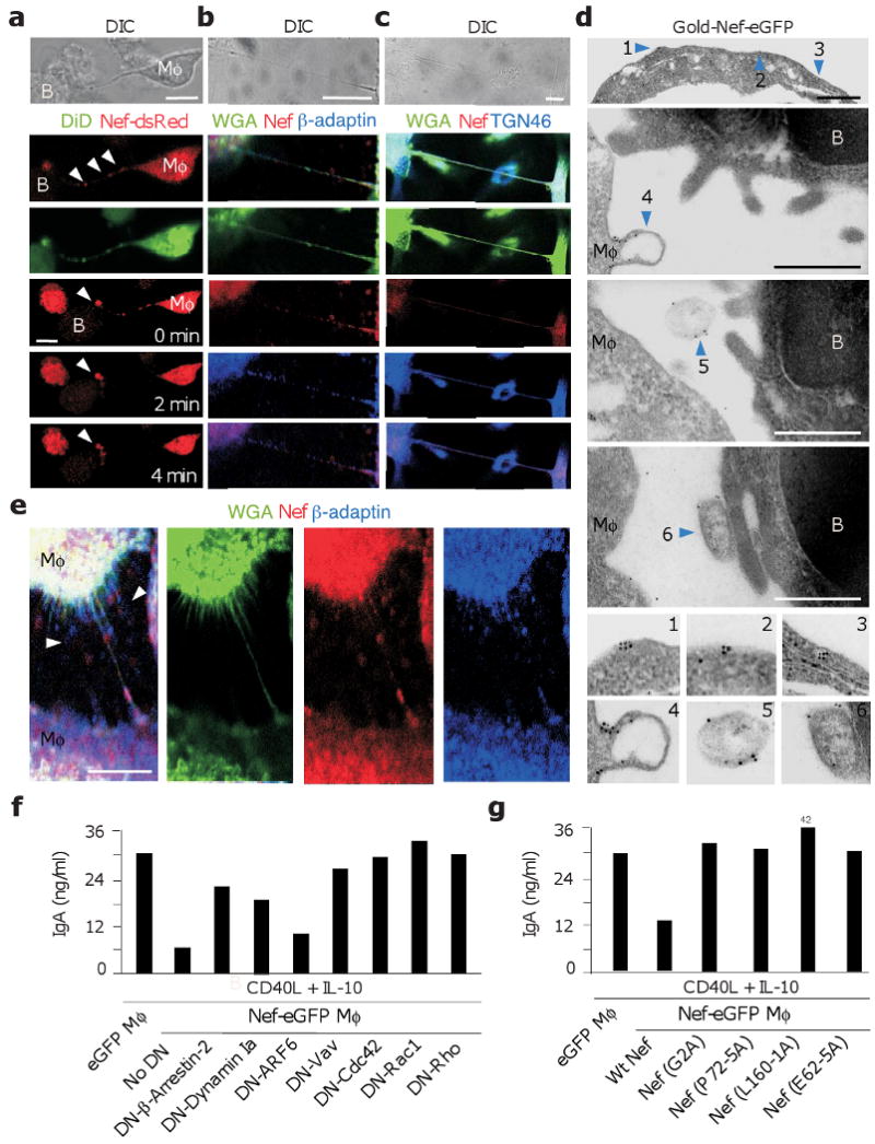
(a) Live DIC (uppermost panels) or confocal imaging of Nef-dsRed-THP-1 macrophage-like cells (Mφ, red) co-stained with the membrane-specific probe DiD (green) and incubated with unstained IgD+ B cells. Arrowheads point to organelle-like beads within an intercellular conduit. The bottom three panels from the top show intercellular transfer of Nef-dsRed over time. Arrowheads point to site of Nef-dsRed accumulation in a B cell. Original magnification, ×40. (b, c) Confocal imaging of ADA-infected primary macrophages (Mφ) stained for Nef (red) and β-adaptin or TGN46 (blue) in the presence of WGA (green). (d) Transmission electron microscopy of Nef-eGFP-THP-1 macrophage-like cells (Mφ) co-cultured with IgD+ B cells for 6 hours and stained with a gold-labeled monoclonal antibody to GFP. Arrowheads 1 and 2, membrane-bound Nef on a Mφ-derived conduit; arrowhead 3, Nef within vesicular and tubular structures from a Mφ-derived conduit; arrowheads 4-6, Nef associated with Mφ budding as well as free or B cell-docked exosome-like bodies, respectively. Original magnification, ×36000. (e) Confocal imaging of ADA-infected primary macrophages stained for Nef (red) or β-adaptin (blue) in the presence of WGA (green). Arrowheads point to extracellular bodies. (f) ELISAs of IgA from IgD+ B cells incubated with THP-1-Nef-eGFP cells nucleofected with either an empty plasmid (control) or a DN plasmid to β-arrestin-2, dynamin Ia, ARF6, Vav, Cdc42, Rac1 or Rho. After 48 h, B cells were sorted and cultured for additional 7 days with or without CD40L and IL-10. (g) ELISAs of IgA from IgD+ B cells incubated with THP-1 cells expressing eGFP, wt Nef-eGFP, Nef (G2A)-eGFP, Nef (P72-5A)-eGFP, Nef (L160-1A)-eGFP or Nef (E62-5A)-eGFP. B cells were cultured as in f. Panels a-g show 1 of 4 experiments yielding similar results. Scale bars equal 10 and 1 μm in confocal and transmission electron microscopy images, respectively.