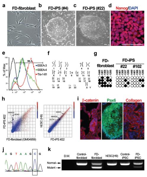
Figure 1. Establishment of FD-iPSCs from patient fibroblasts.
(a-b) FD patient fibroblasts (a) were converted into FD-iPSCs (b, c) following lentiviral transduction with OCT4, SOX2, KLF4, and cMYC. d, Nanog protein expression in FD-iPSC line. e, Flow-cytometry analysis of FD-iPSCs for pluripotent surface markers. f, Karyotype analysis of FD-iPSCs. g, Bisulfite sequencing analysis of NANOG promoter in FD-fibroblast and FD-iPSC clones. h, Global gene expression patterns were compared among FD-fibroblast, FD-iPSCs and human ESCs. i, Teratoma from FD-iPSCs showed three germ-layer differentiation as illustrated by the presence of endodermal epithelia expressing β-catenin, Pax6+ neuroectodermal precursors and mesodermal collagen+ cells. j, Sequencing result showed the 2507+6T>C mutation of IKBKAP in FD-iPSC. k, Analysis of IKBKAP RT-PCR products in mRNA derived from normal and FD-specific fibroblasts and pluripotent stem cells. All scale bars correspond to 50μm.

