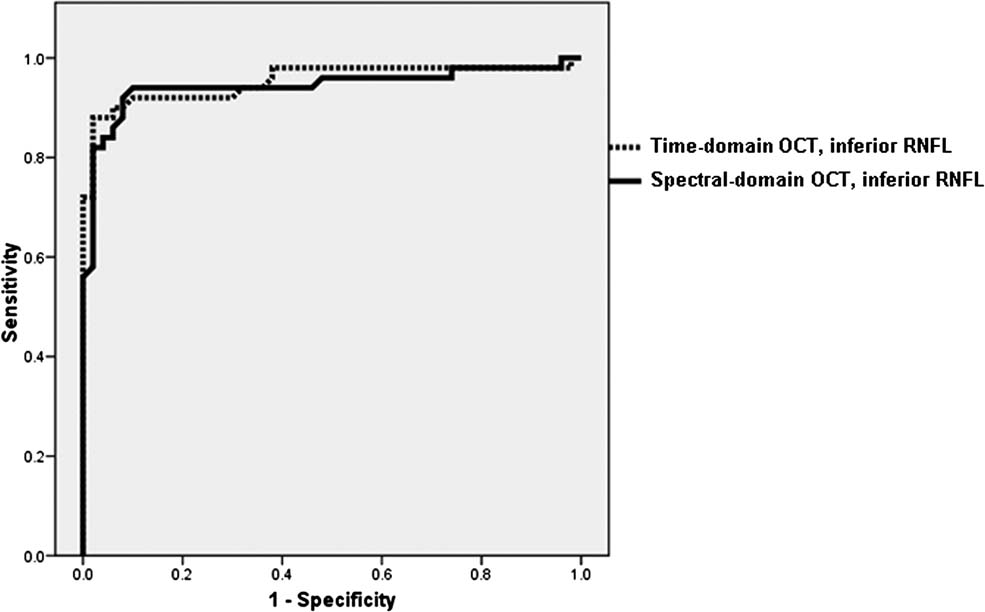
Figure 2.
demonstrates the area under the receiver operator characteristic curves (AUROC) for the best parameter obtained using time-domain optical coherence tomography (inferior RNFL thickness, AUROC = 0.95) and Fourier-domain optical coherence tomography (inferior RNFL thickness, AUROC = 0.94, p = 0.45).

