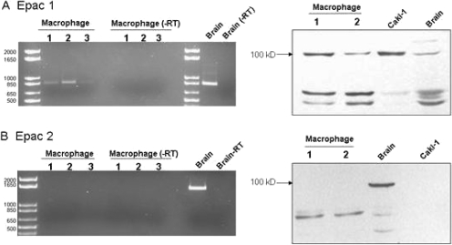
Fig. 4.
Detection of Epac-1 (A), but not Epac-2 (B), in murine macrophages by RT-PCR (left) and Western immunoblotting (right). RT-PCR reactions conducted with and without reverse transcriptase (−RT) were electrophoresed through 1% agarose gels containing ethidium bromide. Reactions were performed with total RNA isolated from three different macrophage isolations (lanes 1–3) and with mouse brain tissue as a positive control. Expected band sizes were 868 base pairs for Epac-1 and 1742 for Epac-2. Western blot analysis was performed with protein lysates from macrophages (lanes 1 and 2) and from mouse brain and Caki-1 cells as positive controls. Epac-1 and Epac-2 migrate as ∼110-kilobase proteins.

