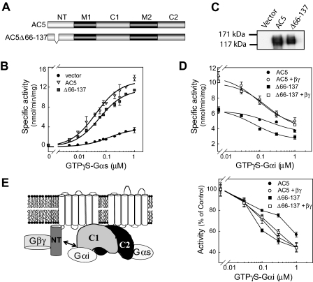
Fig. 4.
Regulation of AC5Δ66–137 by exogenously added GTPγS-Gαs, Gβγ, and GTPγS-Gαi. A, diagram of AC5 and AC5Δ66–137. B, dose response of GTPγS-Gαs stimulated AC5 and AC5Δ66–137 activity. Membranes from HEK293 cells expressing vector, AC5, or AC5Δ66–137 were stimulated with the indicated concentrations GTPγS-Gαs. C, characterization of AC5 and AC5Δ66–137 expression by Western blotting in Sf9 membranes. D, stimulation of Sf9 membranes expressing AC5 or AC5Δ66–137 by 30 nM GTPγS-Gαs in the presence or absence of 100 nM Gβ1γ2 and the indicated concentrations of GTPγS-Gαi. Bottom, each curve was normalized to the AC activity in the absence of Gαi. E, model of Gαi regulation by 5NT. GTPγS-Gαs binds to 5C2 to stimulate activity, whereas GTPγS-Gαs binds to 5C1 to inhibit AC5. 5NT also interacts with 5C1 to limit Gαi inhibition. Addition of Gβγ increases activity and relieves the influence of 5NT on Gαi inhibition. Note that although Gβγ is shown bound to 5NT, it clearly must have additional unknown activation sites.

